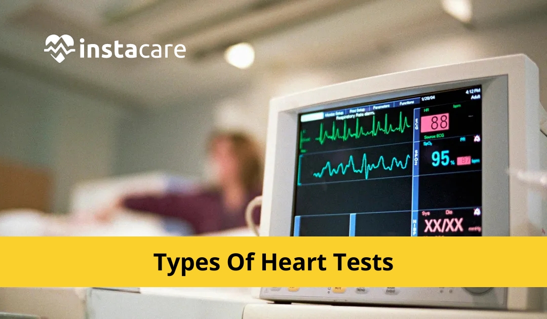
The health of the heart is always paramount and ways and
means to diagnose diseases affecting it or observe its functioning is
offered. Knowledge of the varieties of heart tests may help in the early
diagnosis of the issues and their further control. Here, the ABCs of the more
well-known heart disease tests are presented in terms of their uses and
methods.
Here Are the Types of Heart Tests
1- Angiogram
An angiogram is an X-ray test that is specifically utilized
for the display of blood vessels in the heart. This test entails the use of a contrast
dye to be administered to the coronary arteries by placing a catheter through a
blood vessel in the groin or arm. It is during this procedure that the dye
highlights arteries on the X-ray pictures and thus, helps the physician note
any form of blockage or obstruction on the arteries. Coronary angiography is
frequently used also for managing patients with certain kinds of coronary
artery disease and for performing angioplasty or coronary artery bypass
surgery.
2- Blood Tests
Therefore, related blood tests can provide a lot about the
heart health. For example there is lipid profile test with cholesterol, other
tests include high-sensitivity C - reactive protein (hs-CRP) and troponins that
evaluate inflammation and heart muscle respectively. Increases in the
concentration of enzymes and proteins in the blood provide evidence of the
occurrence of a heart attack or other heart diseases, which means that blood
tests are essential for the identification of signs of a problem at an early
stage.
3- Cardiac Catheterization
Coronary angiography is a non-surgery in the conclusion of
issues with the heart and its blood supply. An extremely slim flexible pipe
referred to as Catheter is passed through a blood vein and is directed straight
to the heart. Strain and oxygen levels in different fragments of the heart,
vein capability and heart valve capability, and illness in the heart walls are
undeniably assessed by this test. As with most other forms of diagnostic
visualizations, it is frequently employed in conjunction with an angiogram.
4- CT Coronary Angiography
CT Coronary Angiography is an indicative procedure which
includes utilization of a CT scanner to produce photos of the coronary
corridors. In anticipation of the sweep, the patient is given a differentiation
color that is controlled through a vein in the patient's body; this assists
with making the corridors more noticeable in the pictures that are taken.
Reverberation, particularly when utilized in blend with
CTCA, is especially important for what, for instance, coronary vein illness has
capabilities that can anticipate blockages or limiting in the coronary conduits
and is a decent device for assessing the gamble of coronary corridor sickness.
5- Doppler Ultrasound
A Doppler ultrasound is a harmless technique that utilizes
sound waves to investigate streams of blood through heart and veins. It helps
the specialists in evaluating the state of the valves of the heart and whether
there is flow that is hindered or whether there is a coagulation in the veins.
This test is generally given along with a routine actual echocardiogram to get
a more itemized assessment of the situation with the heart.
6- Echocardiogram
An echocardiogram is a test wherein piercing sound waves are
coordinated towards the heart and its photos are taken. It gives depictions of
the size and state of the heart, the heart compressions and the idea of
constrictions of the heart walls and those that portray the capability of the
valves of the heart. Different Reverberation Types are transthoracic, transesophageal,
and stress echocardiogram.
7- Electrocardiogram (ECG or EKG)
ECG or E K G is a safe symptomatic test that includes the
recording of the heart's electrical action. Anodes that are little are joined
on the chest, arms and legs in order to lead the electrical driving forces
created by the heart while siphoning. The final result of the interaction is
the diagram of the heart cadence where one can decide different arrhythmias and
potential coronary episodes or some other heart conditions.
8- Electrophysiology Study (EPS)
Electrophysiological review or EPS is another particular
test that spotlights on the electrical working of the heart. This cycle
utilizes catheters with terminals that are set straightforwardly into the heart
through the veins in the crotch region or the arm. EPS distinguishes where part
or the heart's all electrical signs are unpredictable and helps with the
administration of these abnormalities. This test also helps determine what
steps need to be taken; like pacemaker and catheter ablation.
10- Exercise Stress Test
Practice pressure test, or treadmill test, or exercise
ECG-assessment decides the capacity of the heart when in real life. In the test
the patient activities on a treadmill or activities on a bicycle while having
the pulse, circulatory strain and ECG recorded. This test is utilized to
distinguish the seriousness of obstructed veins that supply the heart, judge
the patient's capacity to exercise and gauge the proficiency of treatment in
heart illnesses.
11- Holter Monitor
A Holter screen is characterized as a versatile gadget that
gives the heart's nonstop electrical action for 24 to 48 hours. The patient
gets fitted with the monitor while they carry out their routine activities,
thus offering extended observations of the heart rhythm. Holter monitors are
applied to search for the arrhythmias that are not detected during the standard
ECG and other symptoms such as palpitations. It is particularly useful in the
determination of arrhythmia and in tracking the efficiency of the therapy
applied.
12- Magnetic Resonance Imaging (MRI)
Cardiovascular Alluring Resonation Imaging (X-beam) is an
innocuous imaging evaluation, which is finished including magnets and Radio
waves to take photos of the heart and the veins. It produces capable and clear
nuances on the organ's plan, working, and course to perhaps recognize and show
different heart issues like natural heart infirmity, cardiomyopathy, valvular
ailments, and that is only the start.
13- Myocardial Perfusion Imaging (MPI)
Myocardial Perfusion Imaging (MPI) is an embellishment of
atomic medication that evaluates the blood supply of the myocardium. The
technique incorporates regulating a little portion of radioactive tracer
through the circulatory system and noticing the heart working with the
assistance of a devoted camera. This study commonly involves using MPI during
rest and exercise to evaluate blood flow in the steps.
14- The Positron Emission Tomography (PET) Scan
A PET scan is a refined imaging test that helps give
explicit data in regard to the heart's exhibition and digestion. It is
finished just barely of radioactive tracer into the circulation system and
afterward has a camera that requires some investment photos of the heart. In
particular, PET sweeps are useful in deciding different conditions of ischemia,
assessing the degree of harm of the heart muscles, and assessing the progress
of medicines for heart sicknesses.
Final Thoughts
This information on the different tests for the heart
assists the patients and the medical professionals in arriving at the right decisions
regarding the diagnosis of heart ailments. Ranging from simple imaging
studies to more complex tests, all such tests are helpful and offer essential
information in managing patients’ conditions and ultimately, heart disease. The
next logical step is to monitor one’s health regularly and conduct tests in
case of any previous incidents to avoid declaring war on the heart.
Please book an appointment with the best Cardiologist in Lahore, Karachi, Islamabad, and all major cities of Pakistan through Instacare, or call our helpline at 03171777509 to find a verified doctor for your disease.


